
.jpg)


.jpg)


![[VIDEO] Ultrasound-Guided Injection for Trigger Finger](https://drjordijimenez.com/imagen/100/100/Imagenes/infiltracion-ecoguidada-dedo-resorte-drjordijimenez.jpg)
- Home /
- TREATMENTS /
- ULTRASOUND-GUIDED INFILTRATIONS WITH CORTICOSTEROIDS /
- TREATMENT OF LUMBAR FACET SYNDROME: ULTRASOUND-GUIDED FACET JOINT BLOCK
TREATMENT OF LUMBAR FACET SYNDROME: ULTRASOUND-GUIDED FACET JOINT BLOCK
TREATMENT OF LUMBAR FACET SYNDROME WITH ULTRASOUND-GUIDED LUMBAR FACET BLOCK
DR. JORDI JIMÉNEZ
.jpg)
What is lumbar facet syndrome?
Low back pain is one of the leading causes of medical consultations and sick leave in our country. It is often multifactorial, originating in different structures: muscular, tendon, disc, or even bone. One of these anatomical structures that cause low back pain is the lumbar facet joint, or lumbar facet, located in the posterior part of the lumbar spine. These lumbar facets are responsible for between 15 and 46% of chronic low back pain in our country, and their involvement by inflammatory, traumatic, or osteoarthritic causes leads to what is known as lumbar facet syndrome.
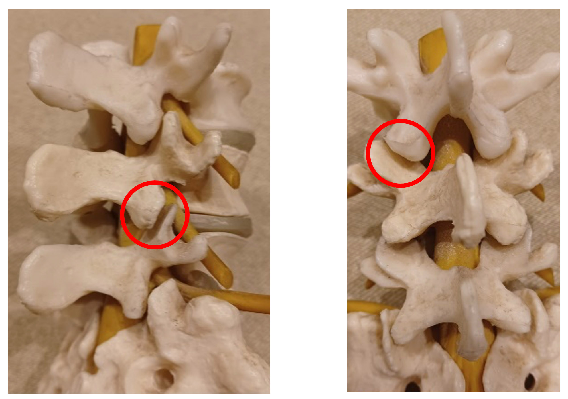
What is the function of the lumbar facets?
The main purpose of these lumbar facets is to provide resistance to rotation and anterior displacement in the spine. They are small and support 15% of the body's total axial load. Therefore, these joints are subjected to 10 times the load per cm² that the knees support.
In prolonged extended postures, the facet joints support a greater axial load, and if we add the previous deterioration of the intervertebral disc, this can reach 70% of the total axial load. Prolonged postures, especially in flexion, can also cause overload and irritation of these joints.
What are the symptoms of lumbar facet syndrome?
- Low back pain radiating to the gluteus, hip, or thigh, mimicking sciatic pain
- Lower back stiffness in the morning.
In the case of facet pain without prior structural impairment, there is the so-called acute locked facet syndrome, in which the patient reports becoming "stuck" when performing a simple movement (picking something up from the floor), presenting with lower back pain and marked reduction in mobility.
How is lumbar facet syndrome diagnosed?
The diagnosis is clinical, based on the patient's history and physical examination. Other structures capable of producing low back pain (vertebral bodies, intervertebral disc, dura mater, nerve roots, sacroiliac joint, muscles, ligaments, and fascia) can overlap the referred pain pattern of facet origin. For this reason, diagnosing the cause of low back pain is sometimes difficult.
How is lumbar facet syndrome treated?
Although initial treatment is symptomatic with local heat, analgesics, and physical therapy, there are chronic cases in which these are not effective, either due to misdiagnosis or concomitant lumbar pathology.
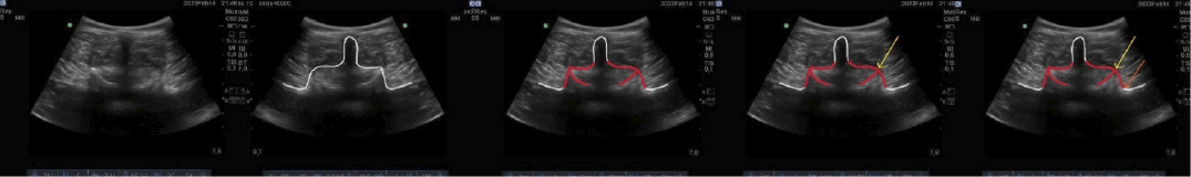
In these cases, ultrasound-guided lumbar facet blocks with anesthetic and corticosteroid offer the possibility of rapid diagnosis and early pain treatment. The anesthetic will block the pain almost immediately, confirming the diagnosis of lumbar facet syndrome. The corticosteroid will treat the inflammation of these facets.
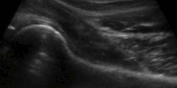
Ultrasound image of the lumbar vertebra: vertebral bone relief in white, lumbar facets in red, yellow arrow: facet block, orange arrow: thermocoagulation (rhizolysis).
What is lumbar rhizolysis?
En casos crónicos en los que los bloqueos son efectivos, pero mejoran al paciente pocas semanas, se emplea la radiofrecuencia, la conocida rizólisis lumbar. Se trata de la termocoagulación de la rama sensitiva que recoge la información dolorosa de estas articulaciones.
Ambos procedimientos ecoguiados pueden realizarse de forma ambulatoria.
Su duración depende de los niveles a los que se practique, pero no excede los 25-30 minutos.
En los casos crónicos, secundarios en la mayor parte a procesos degenerativos (artrosis), la efectividad inicial puede verse reducida a medio plazo siendo necesario repetir la técnica. En cualquier caso , es una técnica segura que proporciona buenos resultados en cuanto a la mejoría del dolor lumbar de origen facetario.
 |
DR. JORDI JIMÉNEZ GENERAL TRAUMATOLOGY SPORTS TRAUMATOLOGY ULTRASOUND-GUIDED THERAPIES |
Do you suffer from lumbar facet syndrome? Make an appointment with Dr. Jordi Jiménez. He will see you in the center of Palma de Mallorca and help you regain your quality of life.
RELATED NEWS
[VIDEO] Ultrasound-Guided Injection for Trigger Finger
Trigger finger is a medical condition in which a finger gets stuck or locked in a bent position and then releases with a characteristic “click.” The condition can be painful and affect a person's ability to move the affected finger. It is more co...
[VIDEO] Ultrasound-guided infiltration of the lumbar facets
This video shows a ultrasound-guided injection of a right L4-L5 lumbar facet. Low back pain is one of the main reasons for medical consultation and work absence in our country. It is often multifactorial, with origins in different structures: muscula...
[VIDEO] Ultrasound-guided infiltration of the hip joint
This video demonstrates an ultrasound-guided hip joint injection, used for the diagnostic and therapeutic management of hip pain. Under real-time ultrasound guidance, the needle is precisely placed in the joint to deliver medication such as local ane...
[VIDEO] Ultrasound-guided infiltration of the proximal sacroiliac joint
This video shows an ultrasound-guided injection of the proximal sacroiliac joint, a procedure used in the diagnostic and therapeutic approach to sacroiliac pain. Under real-time ultrasound guidance, the posterior joint space is precisely accessed to...
[VIDEO] Ultrasound-Guided Interfascial Hydrodissection Middle Trapezius - Rhomboids
Ultrasound-guided interfascial hydrodissection is a minimally invasive technique that involves injecting solution to separate fascial planes, relieve muscle tension, and improve mobility. In this case, it is performed on the Middle Trapezius and Rhom...
[VIDEO] Ultrasound-guided infiltration of De Quervain's tenosynovitis
De Quervain's tenosynovitis is an inflammatory condition affecting the tendons at the base of the thumb in the wrist (Abductor Pollicis Longus [AL] and Extensor Pollicis Brevis [EC], specifically in the first extensor compartment). The affected t...
[VIDEO] Ultrasound-guided infiltration of the acromioclavicular joint
Ultrasound-guided infiltration of the acromioclavicular joint is an interventional procedure that, through ultrasound guidance, allows the administration of analgesic and anti-inflammatory medication directly into the shoulder joint. This joint, form...
[VIDEO] Ultrasound-guided injection for rhizarthrosis
Thumb osteoarthritis is a degenerative disease of the trapeziometacarpal joint. As the severity increases, deformity, pain, and loss of function also increase. Rehabilitation plays an important role in pain control and functional improvement, but whe...
Ultrasound-guided glenohumeral infiltration
What is Ultrasound-Guided Glenohumeral Joint Infiltration? Ultrasound-guided injection of the glenohumeral joint is an interventional procedure that uses ultrasound guidance to infuse analgesic and anti-inflammatory medication into the glenohumeral j...
Ultrasound-guided acromioclavicular infiltration
What is Ultrasound-Guided Acromioclavicular Injection? Ultrasound-guided acromioclavicular injection is an interventional procedure that uses ultrasound guidance to inject analgesic and anti-inflammatory medication into the acromioclavicular joint of...
Ultrasound-guided infiltration of the long head of the biceps
What is Ultrasound-Guided Infiltration of the Long Head of the Biceps? Ultrasound-guided infiltration of the long head of the biceps is an interventional procedure that uses ultrasound guidance to deliver analgesic and anti-inflammatory medication in...
Ultrasound-guided infiltration of the Iliopsoas complex
What is ultrasound-guided injection of the iliopsoas complex? Ultrasound-guided injection of the iliopsoas complex is an interventional procedure that uses ultrasound guidance to deliver analgesic and anti-inflammatory medication to the musculotendin...
Ultrasound-guided interfascial infiltration of the upper trapezius and levator scapulae
What is Ultrasound-Guided Interfascial Infiltration of the Upper Trapezius and Levator Scapulae? [VIDEO] Ultrasound-Guided Interfascial Injection of the Upper Trapezius and Levator Scapulae Ultrasound-guided interfascial injection of the upper trapez...
Interfascial ultrasound-guided infiltration of the lower trapezius and spinal extensor muscles
What is Interfascial Ultrasound-Guided Injection of the Lower Trapezius and Spine Extensor Muscles? Interfascial ultrasound-guided injection between the lower trapezius and spinal extensor muscles is a procedure in which we inject a corticosteroid an...
Interfascial ultrasound-guided infiltration of the middle trapezius and rhomboids
What is ultrasound-guided interfascial injection between the middle trapezius and rhomboids? Ultrasound-guided interfascial injection between the middle trapezius and rhomboids is an interventional procedure that uses ultrasound guidance to place an...
Ultrasound-guided infiltration of rhizarthrosis
What is Ultrasound-Guided Infiltration for Rhizarthrosis?[VIDEO] Ultrasound-Guided Infiltration for Rhizarthrosis Ultrasound-guided infiltration for rhizarthrosis is a procedure that uses ultrasound guidance to introduce anti-inflammatory medication...
Ultrasound-guided infiltration in trigger finger
What is ultrasound-guided injection of trigger finger? This is an ultrasound-guided procedure in which we inject an anti-inflammatory medication around the affected flexor tendon and the obstructing pulley. Performing this injection requires ultrasou...
Ultrasound-guided infiltration of the subacromial bursa of the shoulder
What is Ultrasound-Guided Subacromial Bursa Injection? [VIDEO] Ultrasound-guided injection of the subacromial bursa of the shoulder is a treatment in which an anti-inflammatory medication is injected into the subacromiosubdeltoid bursa of the shoulde...
[VIDEO] Ultrasound-guided injection of the subacromial bursa of the shoulder
Patient with right shoulder bursitis. This video shows the needle approaching the target structure—the subacromial bursa—with the bevel facing down, followed by the injection of corticosteroid and anesthetic solution into the subacromial-subdeltoid b...
Ultrasound-guided hip infiltration
What is an ultrasound-guided hip injection? An ultrasound-guided hip injection is a medical procedure used to relieve pain and inflammation in the hip. It involves injecting a medication (local anesthetic, corticosteroid, hyaluronic acid, or PRP) int...
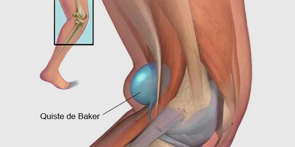
[VIDEO] Ultrasound-Guided Drainage of Baker's Cyst
A Baker's cyst is an accumulation of fluid in the bursa located at the back of the knee. It occurs when joint fluid flows into the bursa but cannot return to the joint, accumulating in the bursa. Baker's cysts are common in older adults or th...
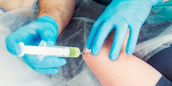
Infiltrations and ultrasound-guided interventions
What is an ultrasound-guided injection? An ultrasound-guided injection is a medical technique in which ultrasound is used to guide the injection of medication into a specific joint or tissue. This technique allows the physician to see the position of...
Epicondylitis and PRP
What is epicondylitis or tennis elbow? Lateral epicondylitis, commonly known as tennis elbow, is considered one of the most prevalent elbow conditions. It is a degenerative tendon process that affects the common extensor tendon, and more specifically...
Plantar fasciitis
What is plantar fasciitis or fasciosis? Plantar fasciitis is a degenerative process that causes inflammation of the plantar fascia, a structure of elastic connective tissue that connects the calcaneus to the metatarsal area. This inflammation causes...
Percutaneous treatment of calcific tendonitis (Barbotage Technique)
What is the barbotage technique? Barbotage is an ultrasound-guided technique that allows for the drainage of calcific material from calcific rotator cuff tendinopathies. There are different percutaneous techniques for the treatment of calcific tendon...
Viscosupplementation (hyaluronic acid)
Viscosupplementation is the intra-articular application of viscoelastic substances to improve synovial fluid or replace it with a higher-quality fluid. Viscosupplementation is used as an alternative treatment in the non-surgical management of osteoar...
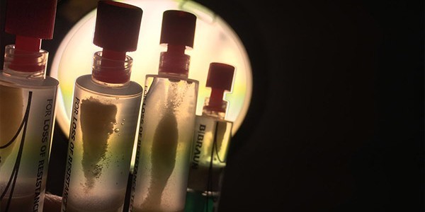
Platelet-rich plasma (PRP) treatment
Platelet-rich plasma (PRP) treatment involves the application of plasma, extracted from the patient, with a platelet concentration well above normal. The purpose of this treatment is to stimulate or trigger the regeneration, scarring, or healing proc...
GENERAL TRAUMATOLOGY
SPORTS TRAUMATOLOGY
ULTRASOUND-GUIDED THERAPIES
IN PALMA DE MALLORCA
BLOG: TAKE CARE OF YOUR HEALTH
![[VIDEO] Ultrasound-guided infiltration of the lumbar facets](https://drjordijimenez.com/imagen/100/100/imagenes-pagina/sindrome-facetario-lumbar-drjordijimenez (1).jpg)
![[VIDEO] Ultrasound-guided infiltration of the hip joint](https://drjordijimenez.com/imagen/100/100/Imagenes/valgo-dinamico-rodilla-drjordijimenez.jpg)