Frequently asked questions about cervical osteoarthritis
- 11/06/2025
What is cervical osteoarthritis or cervical spondylosis?
Cervical osteoarthritis, also defined as cervical spondylosis, refers to the set of age-related degenerative changes that occur in the cervical spine. It is a very common condition; in fact, 85% of people over 65 years of age present some degree of disc degeneration, even in asymptomatic patients.
How do I identify the symptoms of cervical osteoarthritis?
The clinical expression of cervical osteoarthritis can be classified primarily into syndromes such as cervicalgia and radiculopathy. Cervicalgia is a pain that can originate in various cervical structures, including the facet (or interapophyseal) joints, which are also susceptible to degenerative changes. Clinical syndromes manifest as:
- Exclusively cervical pain. This is pain located in the cervical region that can radiate to the shoulder or periscapular area, in a non-dermatous distribution. (an area not served by a specific nerve)
- Cervicobrachialgia (neck pain + pain radiating to the arm): Characterized by pain, sensory disturbances, or neurological deficits corresponding to a dermatome (pain secondary to irritation or compression of a specific cervical root), with or without accompanying neck pain.
What causes neck pain due to osteoarthritis?
Cervicalgia is generally due to muscular and ligamentous factors, such as poor posture or local muscle insufficiency. Previous trauma and degenerative changes in the disc, and particularly in the cervical facet joints (also known as facet joints), can be the source of the symptoms, due to the presence of nociceptive nerve fibers in these structures.
How does cervicoarthrosis pinch the cervical nerve?
Cervical radiculopathy is caused by loss of height and disc protrusion, hypertrophy of the ligamentum flavum and facet joints, and The formation of osteophytes. All of these factors produce stenosis of the spinal canal (the canal through which the spinal cord runs) and foraminal (the openings through which the cervical nerve roots exit), as well as impairment of the nerve root's vascular supply. Only inflammation or irritation of the nerve root during compression can produce pain, which is sustained by the release of chemical mediators.
Diagnosis of cervical osteoarthritis: what tests are performed?
Accurate identification of the pain, along with a thorough medical history and clinical examination, is essential to determine the underlying pathology. Pain can be assessed subjectively using the VAS (Visual Analogue Assessment) scale, or using scales that measure the level of disability caused by neck pain, such as the Cervical Disability Index.
How does the doctor detect the symptoms of cervical pain?
Whether the pain is located in the neck and is accompanied by stiffness or not, suggests degenerative cervical changes. Muscle weakness in the posterior cervical region suggests a muscle sprain or strain, or a soft tissue injury. If the pain worsens with extension, and especially with turning the head to one side, it suggests pain originating in the disk. In many cases, it is also accompanied by headache (2.5% of the general population).
How is cervical facet syndrome clinically diagnosed?
Cervical facet syndrome is suspected when pain is primarily located in the cervical or interscapular region and may radiate to the head, shoulders, or arm without following a dermatomal pattern (unlike radiculopathy). Typically, the pain worsens with ipsilateral extension and rotation of the cervical spine and may improve with flexion. Palpation of the affected facet joints often elicits pain. Precise identification can be achieved by diagnostic blockade of the medial branches of the nerves innervating the facet.
Imaging tests to diagnose osteoarthritis in the cervical region
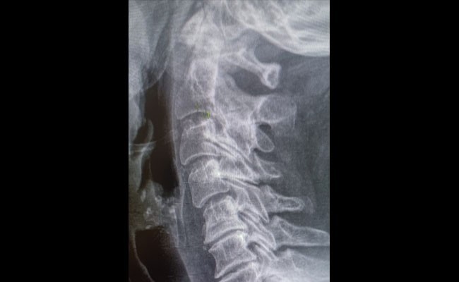
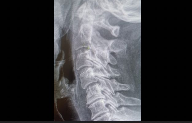
What is seen on a cervical x-ray?
Standard radiographic evaluation of the cervical spine includes anteroposterior, lateral, and oblique views. These should be performed in the standing position whenever possible. Degenerative changes such as intervertebral disc space impingement, osteoarthritis of the uncovertebral joints and facets, the presence of osteophytes, and subchondral sclerosis beneath the vertebral endplates are very common findings in the asymptomatic adult population that are not diagnostic.
When is a cervical CT scan necessary?
Cervical computed tomography (CT) allows visualization of the vertebral bodies and posterior arch in the horizontal (axial) plane, assesses the size and configuration of the spinal contents, evaluates the paravertebral soft tissues, and allows for multiplanar reconstruction, particularly useful in spinal trauma. Its main limitation is inadequate visualization of the contents of the spinal canal.
Why is cervical MRI the best test?
Cervical magnetic resonance imaging (MRI) is the gold standard for evaluating soft tissue in the cervical spine (nerve elements, disc, joint capsule, and ligaments). It is a noninvasive and non-ionizing technique, but it has disadvantages such as the inability to fully distinguish between soft tissue and bone osteophytes, or to demonstrate the presence of posterior element fractures, as well as its susceptibility to movement and metal artifacts.
Who needs electromyography?
Electromyographic studies allow for a differential diagnosis between radiculopathy, compression syndromes, or peripheral neuropathy. Somatosensory and motor evoked potentials and electromyography are the most commonly used.
Conservative treatment for osteoarthritis in the cervical region: what options do I have?
The main goals are to relieve pain, achieve maximum function, and prevent recurrence.
Conservative treatment is the initial treatment of choice in cases of neck pain and radiculopathy. The success rates for nonsurgical treatment of neck pain and cervical radiculopathy range between 70% and 80%. It is based on rest, immobilization with a soft collar, medical treatment (nonsteroidal anti-inflammatory drugs, corticosteroids, muscle relaxants, and antidepressants), and rehabilitation treatment with physical therapy for 4–6 weeks.(See stretching and strengthening exercises for neck pain)
Cervical tractions, as well as translaminar or transforaminal epidural corticosteroid injections, have only been shown to be beneficial in cases of radiculopathy. For cervical facet syndrome, in addition to general measures, physical therapy focuses on specific mobility and strengthening. Ultrasound-guided intra-articular facet joint injections with corticosteroids and local anesthetics are also a conservative treatment option, especially when pain is persistent and localized in these joints. They provide relief and often serve as a diagnostic and therapeutic tool.
Book an appointment with Dr. Jordi Jiménez. He will see you at the center of Palma de Mallorca and help you regain your quality of life.
![[VIDEO] Ultrasound-Guided Injection for Trigger Finger](https://drjordijimenez.com/imagen/100/100/Imagenes/infiltracion-ecoguidada-dedo-resorte-drjordijimenez.jpg)
![[VIDEO] Ultrasound-guided infiltration of the lumbar facets](https://drjordijimenez.com/imagen/100/100/imagenes-pagina/sindrome-facetario-lumbar-drjordijimenez (1).jpg)
![[VIDEO] Ultrasound-guided infiltration of the hip joint](https://drjordijimenez.com/imagen/100/100/Imagenes/valgo-dinamico-rodilla-drjordijimenez.jpg)